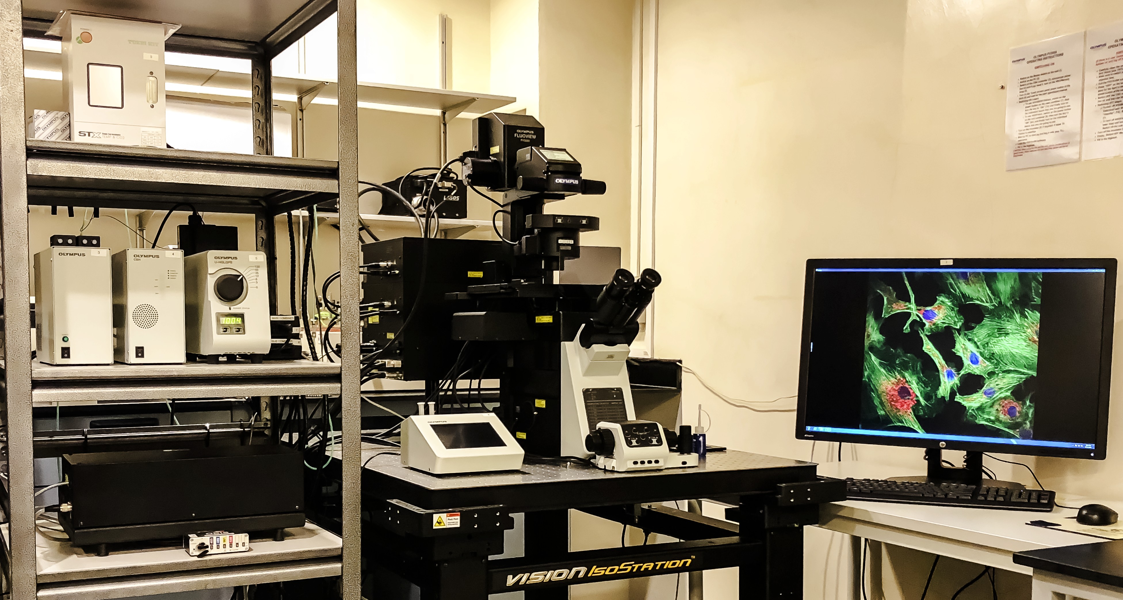
Olympus FV3000 Inverted |
|
|
|
Overview:
- Sensitivity: ★★★★☆
- Speed: ★★★☆☆ (galvano), ★★★★☆ (resonant)
- Detection channels: 2 HSD, 2 PMT, 1 Transmitted Light
- Recommendation: Use if your sample requires higher speed than the Leica SP8 and the sample uses standard fluorophores. This is also the only confocal system with a stagetop incubator for live cell imaging and silicone lens for deep tissue imaging.
Features:
- Diode Lasers: 405, 445, 488, 514, 561, 640nm
- Multidimensional imaging (XYZT)
- Automated Macro to Micro Imaging and stitching of large area of datasets
- Hardware Autofocus
- Simultaneous Spectral Imaging
- 2 Silicone lens for deep tissue imaging and prolong timelapse imaging
- 2 High sensitivity detectors (HSD), 2 PMT detectors and 1 Transmitted Light Detector
- Resonant Scanner: Fast Live Imaging (512 x 512)
- FV31S-SW Acquisition Software
- CellSens for Deconvolution
- Tokai Hit on-stage Incubator (Temperature and CO2 control)
- Other Imaging techniques: FRAP, FRET, Photostimulation, Live Imaging and Spectral Scanning
- Excellent for Live imaging and Deep tissue Imaging
Location: #02-20
Download the instruction manual for this system here
|
| Objective lens |
Magnification/N.A |
Working Distance (μm) |
Immersion Medium |
DIC |
| Olympus PlanApo N |
1.25x/0.04 |
5000 |
Dry |
Yes |
| Olympus UPlanSApo 2 |
10x/0.40 |
3110 |
Dry |
Yes |
| Olympus UPlanSApo |
20x/0.75 |
600 |
Oil |
Yes |
| Olympus UPlanSApo |
30x/1.05 |
800 |
Silicone |
Yes |
| Olympus UApoN 340 2 |
40x/1.35 |
100 |
Oil |
Yes |
| Olympus UPlanSApo |
60x/1.3 |
300 |
Silicone |
Yes |
| Olympus PlanSApo N |
60x/1.42 |
150 |
Oil |
Yes |
| Olympus UPlanSApo |
100x/1.4 |
130 |
Oil |
Yes |
TLL Staff who would like to receive training on this system, please sign up through the Microscopy Training Form.
External users who are interested in this system, please contact the Bioimaging Group 
.png)