Image of the Month
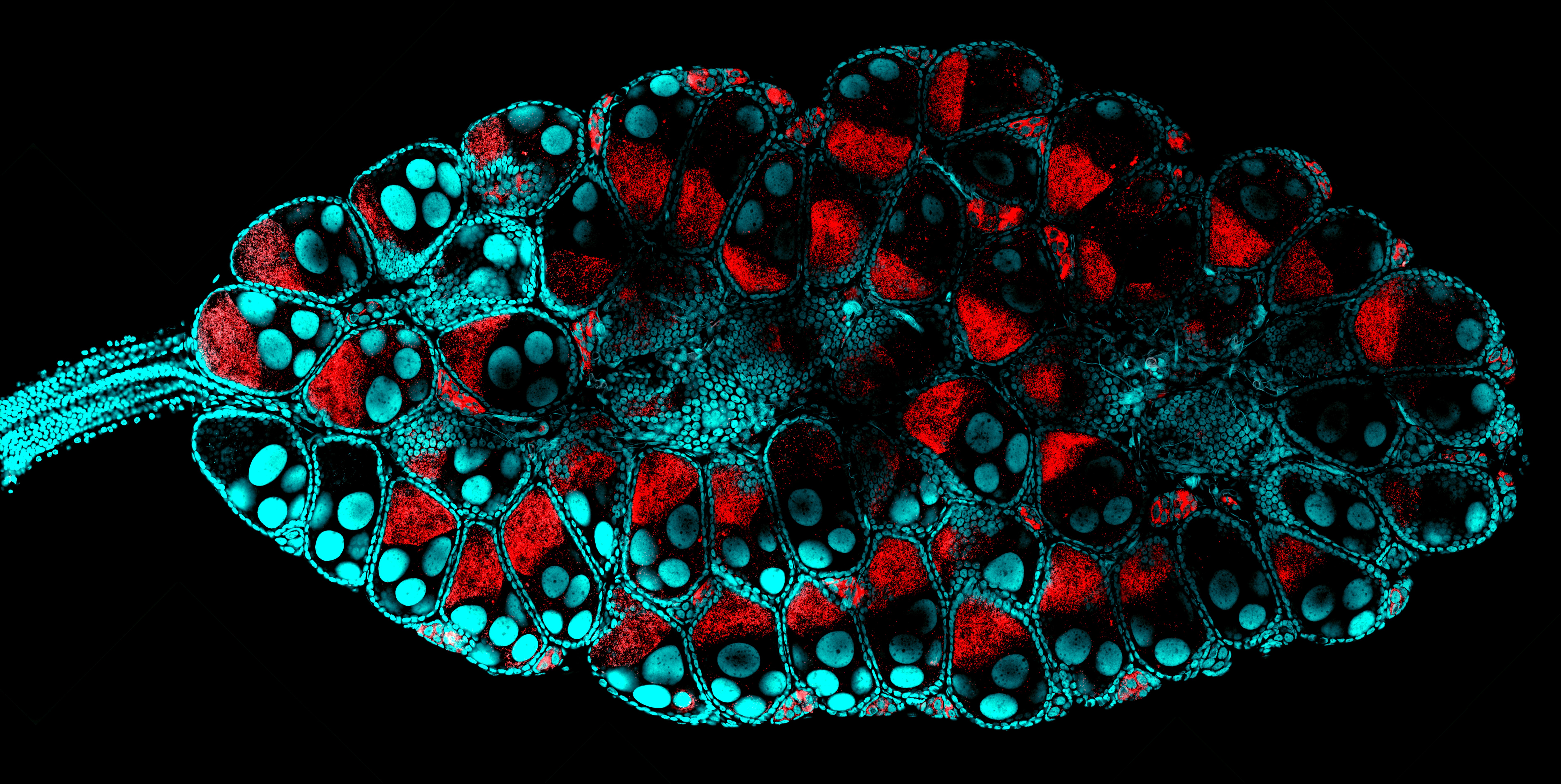
Aedes mosquito ovary infected with Wolbachia bacteria.
The infected mosquito ovary was dissected and labeled with stain for nuclei (blue) and fluorescent probe for Wolbachia (red).
Image acquired on the Olympus FV3000 upright confocal with a 60x Silicone Oil objective.
Taken by
Seah Kwee Boon, Brandon (Cai Yu's lab)
Please submit your most stunning images to the Bioimaging group for the TLL Image of the Month
We welcome images/videos captured using any of the microscopies in TLL, including Stereoscope, Widefield, Confocal and SEM. Please include a brief description in your submission – at least with a title of the image and the microscope/lens used.
Featured submissions from users will win NTUC vouchers worth $40.
.png)