Image of the Month
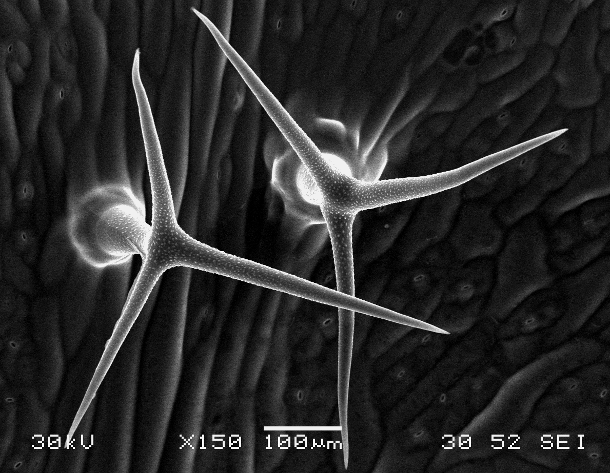
Arabidopsis trichomes (unicellular epidermis hairs) showing branched morphology
Image acquired on the JEOL JSM-6360LV Scanning Electron Microscope at 150× magnification
Taken by Jolly Madathiparambil Saju (Rajani's lab)
.png)
Image acquired on the JEOL JSM-6360LV Scanning Electron Microscope at 150× magnification
Taken by Jolly Madathiparambil Saju (Rajani's lab)
Please submit your most stunning images to the Bioimaging group for the TLL Image of the Month
We welcome images/videos captured using any of the microscopies in TLL, including Stereoscope, Widefield, Confocal and SEM. Please include a brief description in your submission – at least with a title of the image and the microscope/lens used.
Featured submissions from users will win NTUC vouchers worth $40.